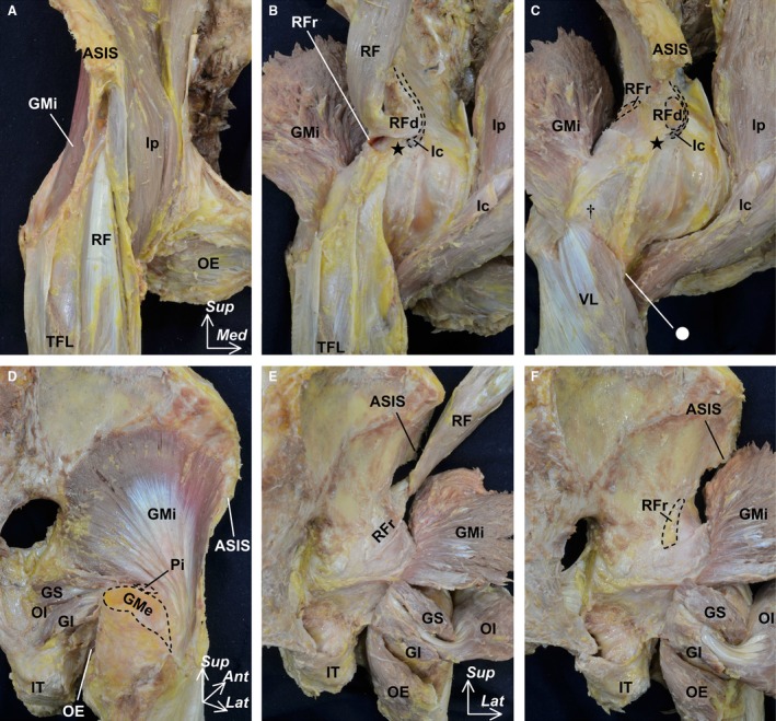
Figure 1.

Macroscopic methods for exposing the outer surface of the hip joint capsule. The pericapsular muscles including the iliopsoas (Ip), gluteus minimus (GMi), rectus femoris (RF), gemelli superior (GS), gemelli inferior (GI), obturator internus (OI) and obturator externus (OE) were reflected from the anterior (A–C) and posterior aspects (D–F). After exposing the surface of the pericapsular muscles (A and D), these muscles were reflected to identify the deep aponeuroses (B and E). In addition, we removed the deep aponeurosis, which did not connect to the outer surface of the joint capsule and the rectus femoris (C and F). Dashed lines indicate the detachment region of the labeled muscle. Circle indicates the inferomedial end of the intertrochanteric line. Dagger indicates the superolateral end of the intertrochanteric line. Star indicates the inferior area of the anterior inferior iliac spine. Ant, anterior; ASIS, anterior superior iliac spine; GMe, gluteus medius; Ic, iliocapsularis; IT, ischial tuberosity; Lat, lateral; Med, medial; Pi, piriformis; RFd, direct head of the RF; RFr, reflected head of the RF; Sup, superior; TFL, tensor fasciae latae; VL, vastus lateralis.
