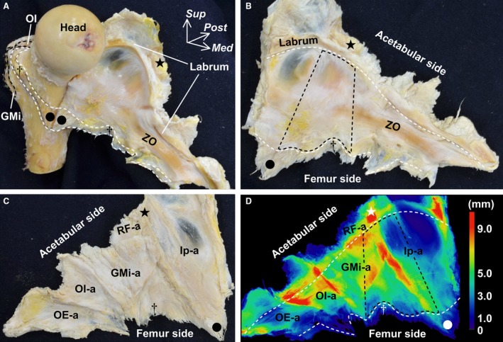
Figure 4.

Appearance of the whole hip joint capsule and its thickness distribution. (A) Anteromedial aspect of the femur. The joint capsule is detached from the femur and reflected medially. White dashed line indicates the distal attachment of the joint capsule. Star indicates the inferior area of the anterior inferior iliac spine. Dagger indicates a tubercle of the femur at the superolateral end of the intertrochanteric line. Circle indicates the inferomedial end of the intertrochanteric line. (B) Inner aspect of the joint capsule. White dashed line on the acetabular side indicates the distal margin of the labrum. White dashed line on the femur side indicates the proximal margin of the capsular attachment. Black dashed area corresponds to the anterosuperior region of the joint capsule surrounded by the star, circle and dagger. (C) Outer aspect of the joint capsule. (D) Local thickness of the same joint capsule analyzed using microcomputed tomography and colored with ImageJ. Color bar represents the approximate thickness corresponding to the different colors. White and black dashed areas correspond to those of (B). GMi, gluteus minimus; GMi‐a, deep aponeurosis of the GMi; Head, head of femur; Ip, deep aponeurosis of the iliopsoas; Med, medial; OE‐a, deep aponeurosis of the obturator externus; OI, obturator internus; OI‐a, deep aponeurosis of the OI, gemelli superior and gemelli inferior; Post, posterior; RF‐a, deep aponeurosis of the rectus femoris; Sup, superior; ZO, zona orbicularis.
