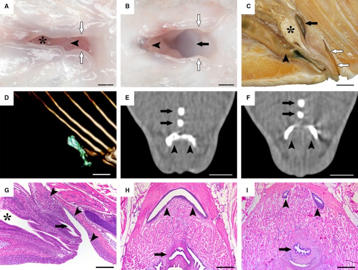
Figure 1.

Morphological investigations of the salmon bursa. (A,B) Ventral view of the salmon cloacal region in a euthanized fish. The asterisk indicates the intestinal aperture in the cranial portion. When pulling the urogenital papilla (arrowheads) cranially and the cloacal labiae (white arrows) aside laterally, the bursa (black arrow) becomes visible. (C) Mediosaggital section of the cloacal region in a formalin‐fixed sample of a male spawning fish, showing the craniocaudal order of the hindgut (arrowhead), the genital tract (black asterisk) and the urinary tract (black arrow) which together forms the urogenital papilla, followed by the bursa (white arrows). (D–F) CT investigations with contrast fluid injected into the bursa. A three‐dimensional render (D) depicts the bursal lumen outline (green) from a lateral view. Note how the bursa (arrowheads) progresses from a crescent shape (E) into two individual blind sacs craniodorsally (F). Black arrows indicate bones of the anal fin. (G–I) Histological examination of the bursa (arrowheads) in mediosaggital (G) and horizontal (H, I) planes, reflecting that of the macroscopic (C) and computed tomography investigations (D–F), while also displaying a prominent lymphoepithelium. Black asterisk indicates the hindgut and black arrows indicate the urogenital tracts. Scale bars, 1 mm (A,B), 1 cm (C–F), 100 µm (G), 200 µm (H,I).
