
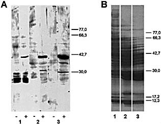
Figure 3.
This article is being made freely available through PubMed Central as part of the COVID-19 public health emergency response. It can be used for unrestricted research re-use and analysis in any form or by any means with acknowledgement of the original source, for the duration of the public health emergency.
Western blot analysis (A) and Coomasie blue staining (B) of different mouse liver fractions. According with Sect. 4 and Fig. 2: liver supernatant obtained after 100,000×g centrifugation (lane 1), DEAE‐cellulose (lane 2) or Sephadex G‐100 chromatography (lane 3). SDS‐PAGE was performed in 14×16 cm gels using a gradient of 10‐12.5% of polyacrylamide. Transferred proteins were incubated with serum from non‐infected (‐) or MHV‐infected (+) mice. The positions of molecular mass markers (kDa) are shown at right.