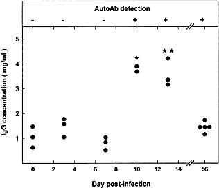
Figure 6.

This article is being made freely available through PubMed Central as part of the COVID-19 public health emergency response. It can be used for unrestricted research re-use and analysis in any form or by any means with acknowledgement of the original source, for the duration of the public health emergency.
Time course of Ab to the 40‐kDa liver protein and plasma Ig level in MHV‐infected CBA/Ht mice. Upper panel: Western‐blot reactivity of serum from MHV‐infected CBA/Ht with the 40‐kDa liver protein (see Fig. 1 for details); (+) means that serum from all animals tested reacted with the mouse liver 40‐kDa protein; (–) means absence of reactivity. Lower panel: Total IgG concentration of the corresponding sera was determined as indicated in Sect. 4. Values correspond to individual mice. Results were subject to statistical analysis by using the Mann‐Whitney U‐test. *p<0.1, **p<0.05.
