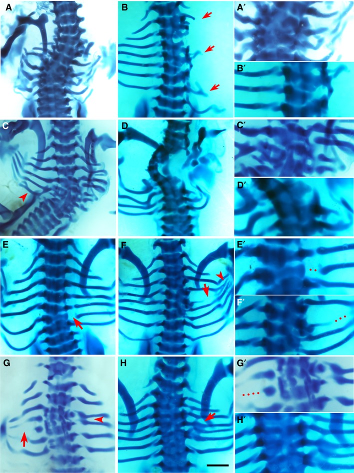
Figure 3.

Phenotypes by bone morphogenic protein (BMP)4. Alcian blue‐stained chick embryos at Hamburger and Hamilton (HH) stage 32‐33, dorsal view. Red arrows and arrowheads indicate the phenotype. The right column (A’–H’) shows higher magnifications of the phenotypic area, with dotted lines indicating missing rib parts. Scale bar, 1 mm. (A) Multiple missing and shortened ribs. Scoliosis is noted, as well as fusion and deformity of vertebrae T3–T7. (B) Missing second, third, fifth and sixth right ribs, with only shortened remnants of the first, fourth and seventh ribs. There is malformation of vertebrae T2–T6. (D) Missing fifth left rib and scoliosis. Fusion and malformation of vertebrae T4–T7. Fusion of left sixth and seventh ribs are indicated by arrowhead. (F) Missing first, second and third ribs bilaterally as well as missing fourth right rib and shortened fifth right rib. There is a hemi vertebral deformity of T1–T5 and ectopic cartilage deposition. (E) Proximal defect of sixth right rib. (F) Vertebral defects and fusion of T4–T6 causing a reduced amount of cartilage deposition in the form of the fifth rib with shaft defect (arrow) and the rib inferior to this, presumably the fifth rib, is then attached to T6 along with the sixth rib. An eighth rib pair is noted. Fusion and branching are also noted at the distal ends (arrowhead). (G) Missing fourth right rib and shortened fifth left rib (arrow). Fusion of the third and fourth ribs on the right (arrowhead). (H) The vertebral column is fused from T1 to T7 and the fifth right rib is separated from its vertebral origin but fused to the proximal end of the fourth rib to create a bifurcated appearance.
