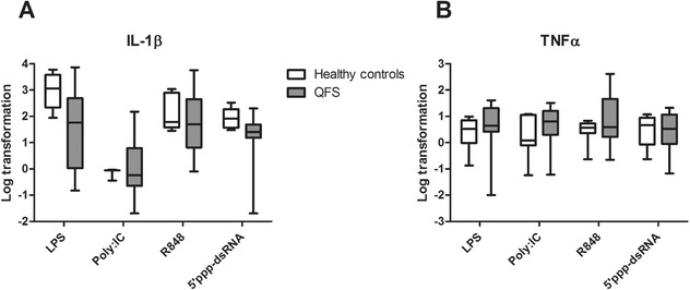
Figure 2.

mRNA expression of IL‐β and TNF‐α in QFS patients compared to healthy controls, Monocytes were stimulated with LPS, Poly I:C, R848, or 5’ppp‐dsRNA for 24 hours. (A) Monocytes of QFS patients showed significantly decreased mRNA expression of IL‐1β compared to healthy controls when stimulated with 5’ppp‐dsRNA (p = 0.036). (B) Monocytes of QFS patients show no significantly difference in mRNA expression of TNF‐α compared to healthy controls. mRNA expression was measured with qPCR. The following primers were used: TNFα FWD‐5’‐AACGGAGCTGAACAATAGGC‐3’, REV‐5’‐TCTCGCCACTGAATAGTAGGG‐3’, IL‐1β FWD‐5’‐GCCCTAAACAGATGAAGTGCTC‐3’, REV‐5’‐GAACCAGCATCTTCCTCAG‐3’. RNA expression was corrected for differences in loading concentration using the signal of housekeeping protein GAPDH. Data were analyzed with the Mann–Whitney test and are depicted as mean ± SEM. Data are derived from one single experiment that consisted of 13 patients and seven healthy controls. Abbreviations: QFS, Q fever fatigue syndrome; Poly I:C, R848, and 5’ppp‐dsRNA, various viral ligands; RPMI, Roswell Park Memorial Institute culture medium; qPCR, quantitative real‐time PCR. *p ≤ 0.05; **p ≤ 0.01
