
Figure 2.
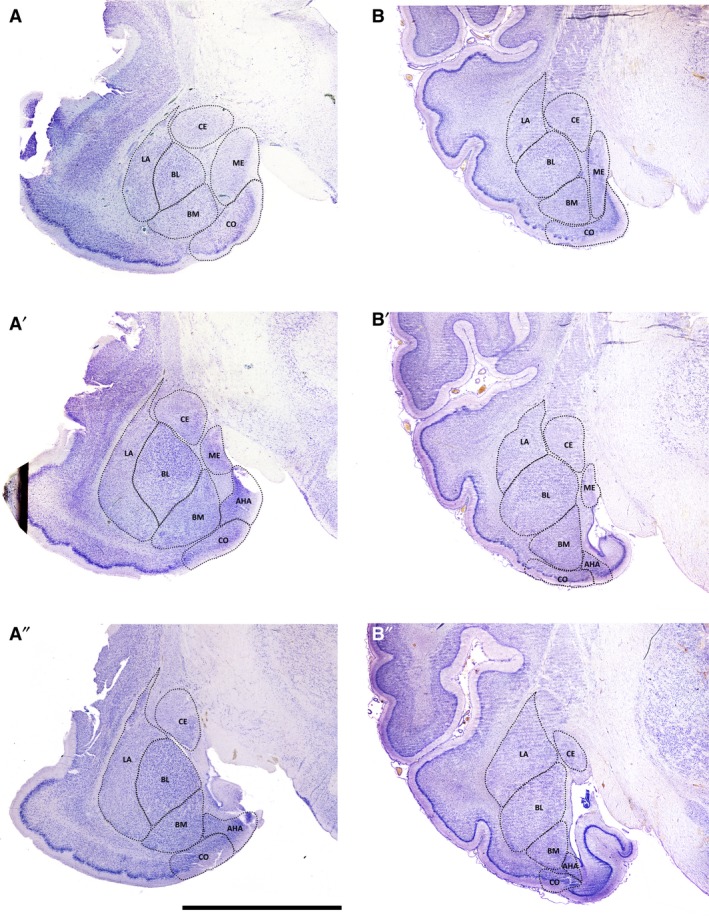
Low‐magnification photomicrographs of representative coronal sections through the fox and pig amygdala, illustrating the locations and boundaries of the main nuclei. A–A″, fox; B–B″, pig. Letter codes represent nuclei as follows: AHA, amygdalo‐hippocampal area; BL, basolateral; BM, basomedial; CE, central; CO, cortical; LA, lateral; ME, medial. Scale bar = 5 mm for A–A″ and 8 mm for B–B″.