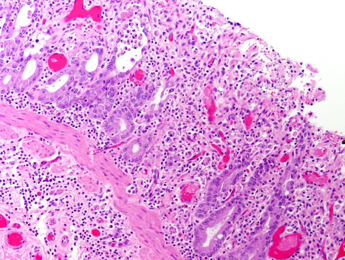
Figure 6.

This illustration shows the H&E stain of jejunum from an adult horse with ECoV infection. There is loss of crypts and few remaining crypts are dilated, lined by attenuated epithelium and contain sloughed necrotic enterocytes. The lamina propria and superficial submucosa are expanded by inflammatory infiltrates. Capillaries and venules in the mucosa and submucosa are occluded by fibrin thrombi.
