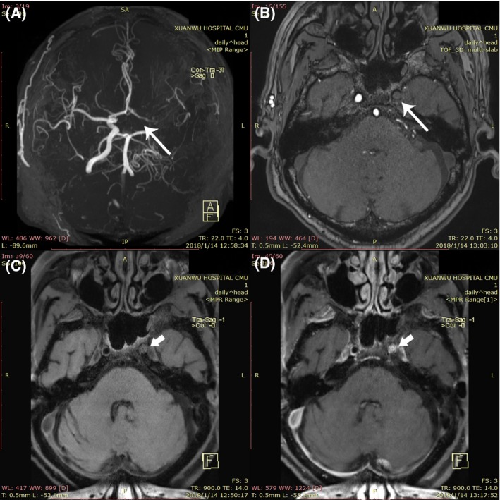
Figure 1.

Features of moyamoya vessels on TOF MRA and HR‐MRI map. TOF MRA shows (A) cross‐sectional image showed moyamoya vessel, (B) the left internal carotid artery with complete occlusion (long arrow), (C) T1‐weighted HR‐MRI, and (D) contrast‐enhanced HR‐MRI present the concentric inward remodeling (short arrow) in the left internal carotid artery lumen
