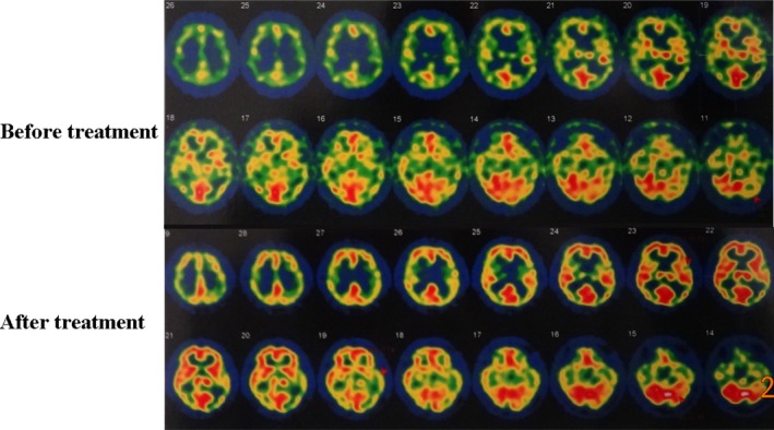
Figure 3.

Perfusion presented on SPECT maps prior to and post‐RIC treatment. SPECT shows that the perfusion decreases in bilateral frontal, parietal, occipital, and temporal lobes before RIC regimen. After 1 year of RIC treatment, the perfusion is significantly enhanced in all lobes
