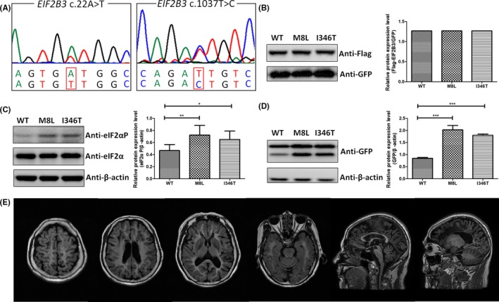
Figure 2.

Functional analysis of variants c.22A>T and c.1037T>C within EIF2B3 and brain MRI of the patient. A, Sequencing chromatograms of variants c.22A>T (p.M8L) and c.1037T>C (p.I346T) within EIF2B3. B, HEK293T cells were cotransfected with p3 × flag‐EIF2B3 (WT or mutant) with pEGFP‐N1 as exogenous control. Neither p.M8L nor p.I346T destabilized the protein appreciably. C, Both p.M8L and p.I346T enhanced eIF2α phosphorylation without increase of total eIF2α. *P < .05 and **P < .01. D, HEK293T cells were cotransfected with p3 × flag‐EIF2B3 (WT, or mutant) with (pEGFP‐ATF4‐5′UTR). After incubated with anti‐GFP antibody, the upper bands reflected the fusion protein containing the product translated from the AUG within ATF4 5′UTR and EGFP, and the lower bands reflected only EGFP, which were regulated by the uORFs in ATF4 5′UTR. Both p.M8L and p.I346T increased the level of expression of EGFP than WT. ***P < .001. E, Brain MRI of proband 4 at her 60 showed extensive white matter hyperintense on FLAIR and markedly hypointense on T1‐weighted images
