
Figure 3.
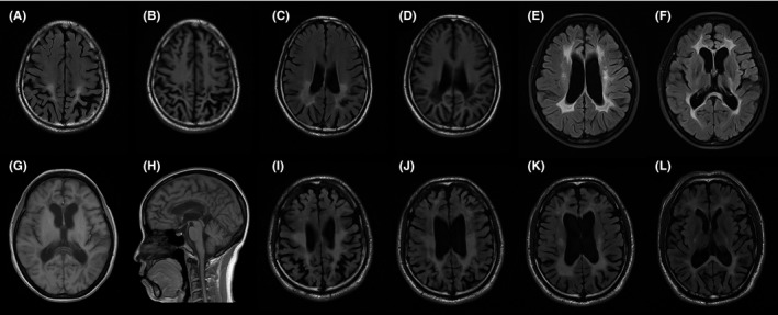
Brain MRI of patients with ALSP. A, B, C and D, MRI of proband 1 at his 43 showed the broadening and deepening of sulus with occipital prominence, and occipital periventricular white matter hyperintense on FLAIR and hypointense on T1‐weighted images. E, F, G and H, MRI of proband 2 at her 46 showed the white matter adjacent to the lateral ventricle hyperintense on FLAIR and hypointense on T1‐weighted images, and thinning of the corpus callosum. I, J, K and L, MRI of proband 3 at his 38 showed widespread white matter hyperintense and obvious cortical atrophy on FLAIR