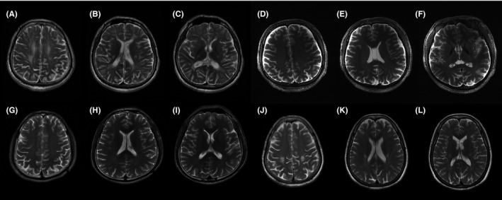
Figure 4.

T2‐weighted MRI on the same three levels (centrum semiovale, corona radiate, and internal capsule) of patients with GLD. A, B and C, MRI of proband 5 at his 21 showed partly confluent hyperintense areas in white matter near the lateral ventricle and posterior limbs of internal capsule. D, E and F, MRI of proband 6 at his 28 showed no visible abnormality. G, H and I, MRI of proband 7 at his 28 showed no visible abnormality. J, K and L, MRI of proband 8 at her 46 showed symmetric hyperintense in bilateral corticospinal tracts
