
Figure 2.
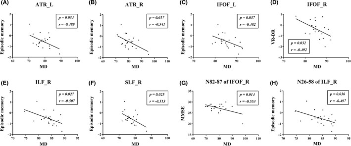
The correlations between MD values and cognition assessment in WMH‐MCI. A‐C and E‐F, Significantly negative correlations between episodic memory and the mean MD of the following tracts: left ATR (r = −.489, P = .034), right ATR (r = −.541, P = .017), left IFOF (r = −.482, P = .037), right ILF (r = −.507, P = .027), and right SLF (r = −.513, P = .025). D, The mean MD of the right IFOF correlated negatively with VR‐DR (r = −.492, P = .032). G, The MD values in the anterior component of the right IFOF (nodes 82‐87) were negatively associated with MMSE (r = −.553, P = .014). H: The right ILF showed significant negative correlation with episodic memory in the posterior and intermediate component (nodes 26‐58) (r = −.497, P = .030). MCI, mild cognitive impairment; WMH, white matter hyperintensities; MD, mean diffusivity; axial diffusivity; ATR_L, left anterior thalamic radiation; ATR_R, right anterior thalamic radiation; IFOF_L, left inferior fronto‐occipital fasciculus; IFOF_R, right inferior fronto‐occipital fasciculus; ILF_R, right inferior longitudinal fasciculus; SLF_R, right superior longitudinal fasciculus; MMSE, Mini Mental State Examination; VR‐DR, Visual Reproduction‐delayed recall