Figure 1.

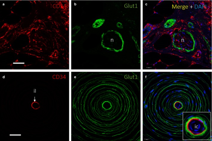
Extraneurial component in peripheral nerve and Pacinian corpuscles. Dual immunofluorescence for (a,c,d,f) CD34 and (b, c, e and f) Glucose transporter 1 (Glut1) showing endoneurium‐derived cells and perineurium‐derived cells, respectively, in a cross‐sectioned cutaneous peripheral nerve (a–c) and a Pacinian corpuscle (d–f). In the peripheral nerve, Glut1 identified the perineurium (b,c), and there was a fine, CD34‐positive network inside of the perineurium that corresponded to the endoneurium (a,c); some connective tissue, with CD34+ fibroblasts, is observed surrounding the nerve, which might correspond to epineurium. Additional findings in the figure are a very faint Glut1 expression corresponding to eccrine ducts (b) and several CD34+ connective tissue structures like blood vessels endothelia, fibroblasts and adipocytes (a,c). In the Pacinian corpuscle, CD34 identified a thin, intermediate layer (d,f), which was surrounded by several outer‐core lamellae (e,f). il, intermediate layer; n, nerve. Scale bar: 60 µm (a–c); 50 µm (d–f); ic, inner core.
