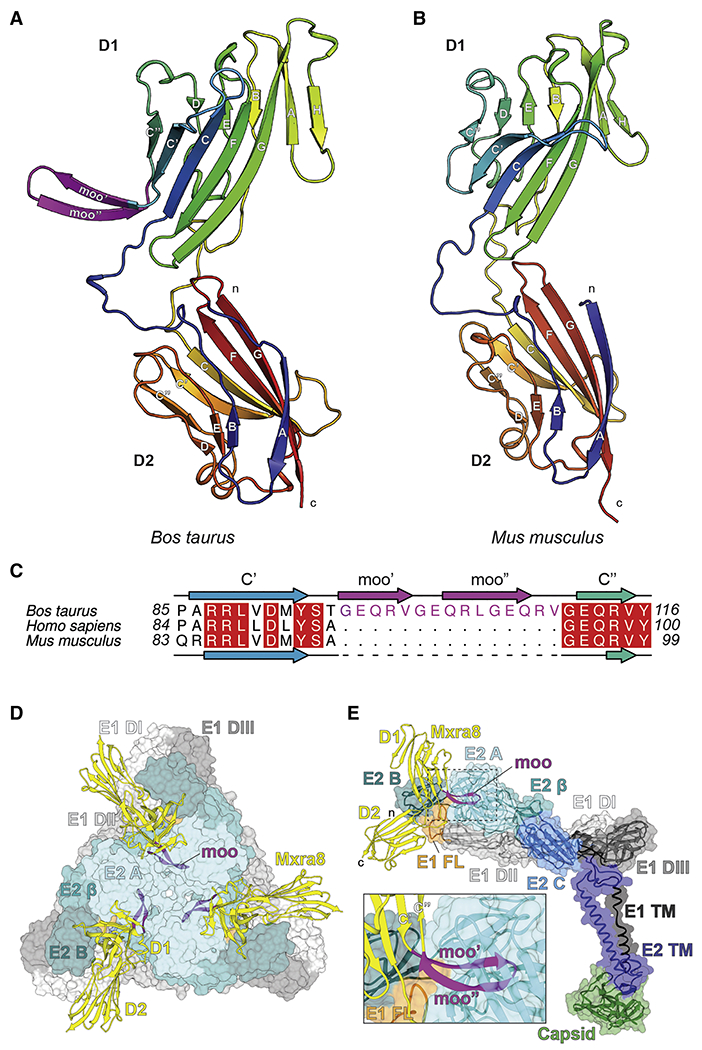
Figure 2. Structure of cattle Mxra8.

A-B. Ribbon models of the (A) cattle and (B) mouse Mxra8 (PDB 6NK3) structures determined by X-ray crystallography. The two Ig-like domains are colored by Jones’ rainbow scheme with the N-terminus in blue and C-terminus in red. The β-strands of each Ig domain are labeled according to standard convention. The N- and C-termini are labeled in lowercase. C. Structure-based alignment of Bos taurus (cattle), Homo sapiens (human), and Mus musculus (mouse) Mxra8 protein sequences highlighting the 15-amino acid insertion site between the C’-C’’ loop in D1. D-E. Docking of cattle Mxra8 onto a cryo-EM model of mouse Mxra8 bound to CHIKV VLPs, viewed from a trimeric spike (D), an E2-E1 subunit (E), and enlarged to highlight the clash (inset). Cattle Mxra8 is colored yellow and labeled by domain, with the moo insert depicted in magenta. The N- and C-termini are labeled in lowercase. Within the inset, the C’, ‘moo’ insert, and C’’ β-strands are labeled, and the E1 fusion loop is depicted in orange. Structural proteins are colored and labeled by domain. E1: DI, light grey; DII, medium grey; DIII, dark grey; fusion loop, orange; TM region, black. E2: A domain, light cyan; β-linker, medium cyan; domain B, dark cyan; domain C, medium blue; TM region, dark blue. Capsid, green. See Fig S2 and S3.
