
Figure 2.
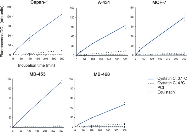
Internalization of cystatin C in cancer cell lines measured by flow cytometry. Five human cancer cell lines were used: MCF‐7, MDA‐MB‐453, MDA‐MB‐468, A‐431 and Capan‐1. Subconfluent cells were incubated in medium containing fluorescence‐labelled protein, either cystatin C, PCI or equistatin. NaCl/Pi was used as control. Cells were incubated for 10 s, 5 min, 30 min, 2 h or 6 h, respectively. Cell fluorescence was measured and median cell fluorescence was calculated, corrected for the control value and then related to the degree of labelling (DOL) of the protein used. The MCF‐7, MDA‐MB‐453 and MDA‐MB‐468 cell lines were in addition incubated with labelled cystatin C at 4 °C as described above. Each of the diagrams shows the results of three independent experiments (A‐431 experiments were carried out twice). The lines are drawn through the average value of the three results at each time point.