
Figure 2.
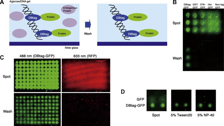
Immobilization and purification of DBtag‐fusion proteins on agarose/DNA microplate. (A) Schematic diagram showing immobilized DBtag‐fusion proteins on the microplate before and after washing. (B) Only DBtag‐GFP was immobilized on the microplate, while washing removed all other tagged GFPs and the untagged GFP. (C) DBtag‐GFP and RFP (untagged) were mixed together and the mixture was spotted on the microplate. The DBtag‐GFP was immobilized on the microplate, while the RFP was washed out and no RFP fluorescence was observed. (D) GFP (untagged, upper spot) and DBtag‐GFP (lower spot) were spotted on the microplate. The DBtag‐GFP was immobilized on the microplate by 5% of detergent treatment (5% Tween‐20 and 5% NP40 panels), while the GFP was washed out and no GFP fluorescence was observed.