
Figure 3.
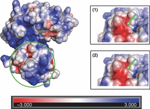
Electrostatic potential distribution of SARS‐CoV helicase: the basic (dark blue coloured) region involved in DNA binding and the dimer interface (circled in green) are shown. On the opposite side of the protein, S259/L regions in the wild‐type (inset 1) and mutated form (inset 2) of the viral enzyme are also shown.