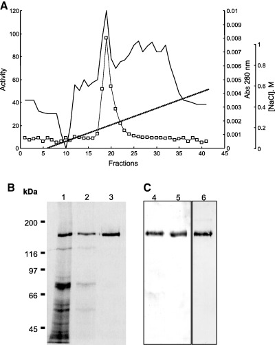
Figure 1.

Chromatographic purification of midgut aminopeptidase from A. pisum. (A) Chromatography on Mono Q equilibrated with 20 mm Tris/HCl buffer (pH 7.0)/0.1% Triton X‐100. Elution was accomplished with a gradient of 0–600 mm NaCl gradient in the same Tris buffer (substrate used LeupNA). (B) SDS/PAGE of samples obtained after the steps from A. pisum APN purification (12% polyacrylamide slab gels, silver staining). Lane 1, midgut homogenate; lane 2, Triton X‐100‐released proteins from midgut cell membranes; lane 3, Mono Q eluate (purified aminopeptidase). (C) Glycoprotein detection (Dig Glycan detection kit), after western blots of proteins. Lane 4, midgut homogenate; lane 5, purified APN; lane 6, purified with the differentiation kit with the mannose‐specific lectin Galanthus nivalis agglutinin.
