
Figure 5.
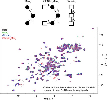
Structural representations of the three glycans investigated in this work. Shown below is the HSQC spectrum of the holo‐CRD (0.4 0.7 mm) at pH 4.2 in the absence (black) and presence (green) of 5 mm Man3 (12.5‐fold molar excess), 10 mm GlcNAc2Man3 (14‐fold molar excess; red), and 10 mm GlcNAc3 (14‐fold molar excess; blue). The addition of Man3 to the CRD at low pH yielded no changes in the CRD spectrum, suggesting that Man binding to the CRD is pH‐dependent. Some small perturbations were observed upon addition of GlcNAc2Man3 and GlcNAc3 at low pH, and suggest that binding of the CRD to the GlcNAc moiety may not be completely abolished at low pH.