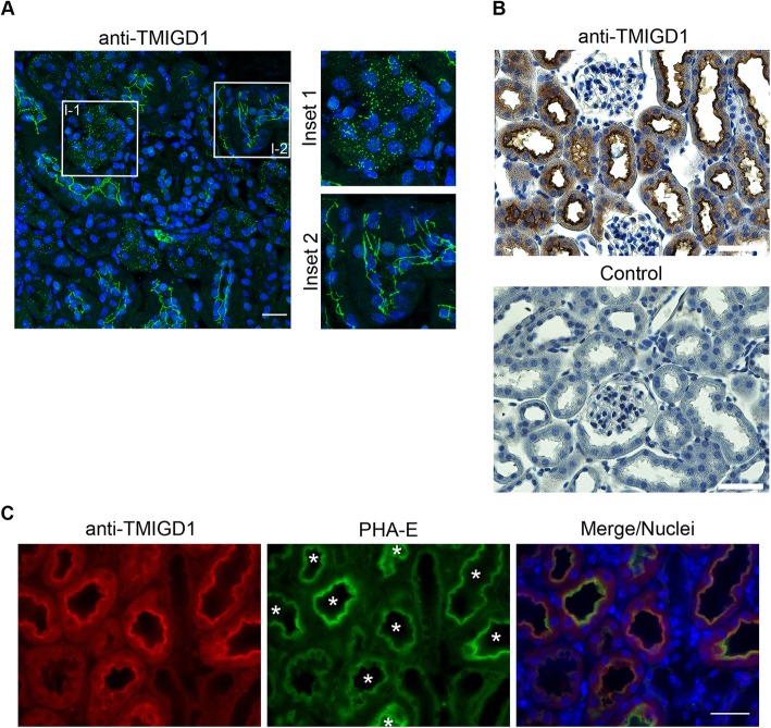
Fig. 2.
TMIGD1 is expressed by renal epithelial cells with distinct subcellular locations. a Immunofluorescence analysis of murine kidney sections with TMIGD1 antibodies (#HPA021946). The insets (Inset 1, Inset 2) show magnifications of the areas demarcated with rectangles (I-1, I-2) in the left panel. Note that TMIGD1 is enriched at cell-cell contacts in some tubular epithelial cells but is localized in the cytoplasm in others. Scale bar: 100 μm. b Immunohistochemistry on murine kidney sections with antibodies against TMIGD1 (#HPA021946). Control stainings (bottom panel) were performed in the absence of the primary antibodies. Note that TMIGD1 is expressed by tubular epithelial cells but is absent from glomerular cells. Scale bars: 50 μm. c Immunofluorescence analysis of murine kidney sections. Kidney sections were stained with FITC-conjugated PHA-E lectin and with anti-TMIGD1 antibodies (#HPA021946). Note that all TMIGD1-positive structures are positive for PHA-E (marked by asterisks) suggesting preferential expression of TMIGD1 by epithelial cells of proximal tubules. Scale bar: 100 μm

