
Fig. 1.—
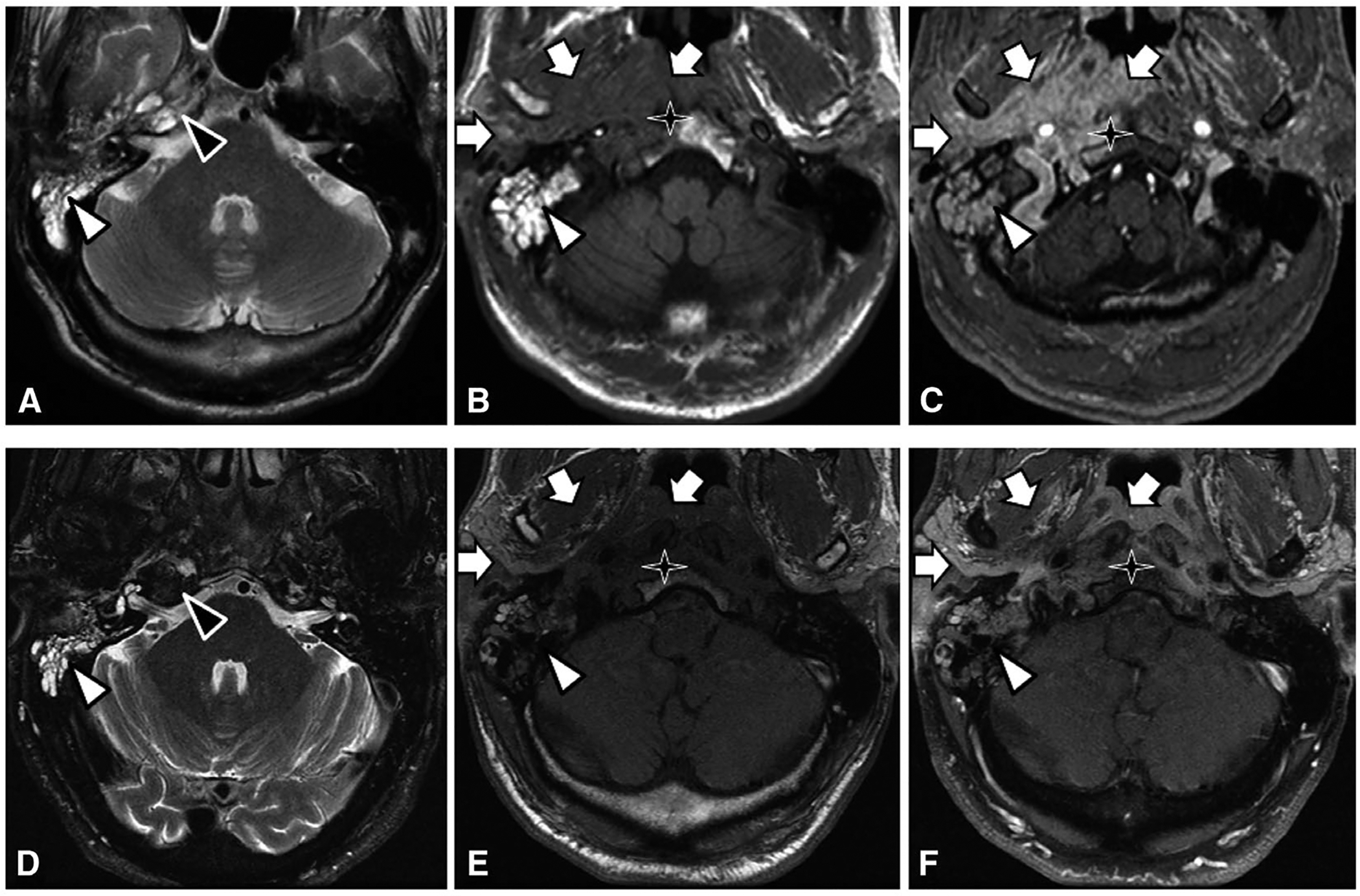
(A-C) Pre-treatment images of right petrous apicitis. T2-weighted image (A) at the level of the petrous apex demonstrating fluid in the pneumatized right petrous apex (black triangle) and mastoid air cells (white triangle). T1 pre-contrast (B) and post-contrast (C) images at the level of the nasopharynx demonstrating abnormal enhancement of the right nasopharynx, parapharyngeal space, and masticator space (white arrows), compatible with infection and inflammation. There is abnormal T1 pre-contrast hypointense marrow signal of the right aspect of the clivus (star), with post-contrast enhancement compatible with osteomyelitis. (D-F) Post-treatment images. T2-weighted image (D) shows resolution of abnormal fluid signal at the right petrous apex. T1 pre- and post-contrast images show near complete resolution of abnormal signal and enhancement of the clivus, right nasopharynx, parapharyngeal space, and masticator space.