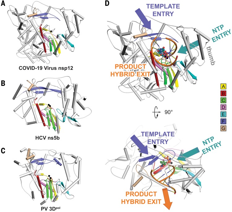
Fig. 3. The RdRp core region.
(A to C) Structural comparison of COVID-19 virus nsp12 (A), HCV ns5b (PDB ID: 4WTG) (13) (B), and PV 3Dpol (PDB ID: 3OLB) (14) (C). The three structures are displayed in the same orientation. The polymerase motifs (motifs A to G) have the same color scheme used in Fig. 1A. (D) The template entry, NTP entry, and product hybrid exit paths in COVID-19 virus nsp12 are labeled in slate, deep teal, and orange colors, respectively. Two catalytic manganese ions (black spheres), pp-sofosbuvir (dark green spheres for carbon atoms), and primer template (orange) from the structure of HCV ns5b in complex pp-sofosbuvir (PDB ID: 4WTG) (13) are superposed to COVID-19 virus nsp12 to indicate the catalytic site and nucleotide binding position.

