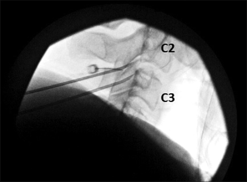
Figure 1.

Radiographic imaging demonstrating a lateral view of the radiofrequency neurotomy of the third occipital nerve using a 16-gauge RFA probe. A third RFA probe was eventually placed between the two probes shown in this image ensuring coagulation of the third occipital nerve
