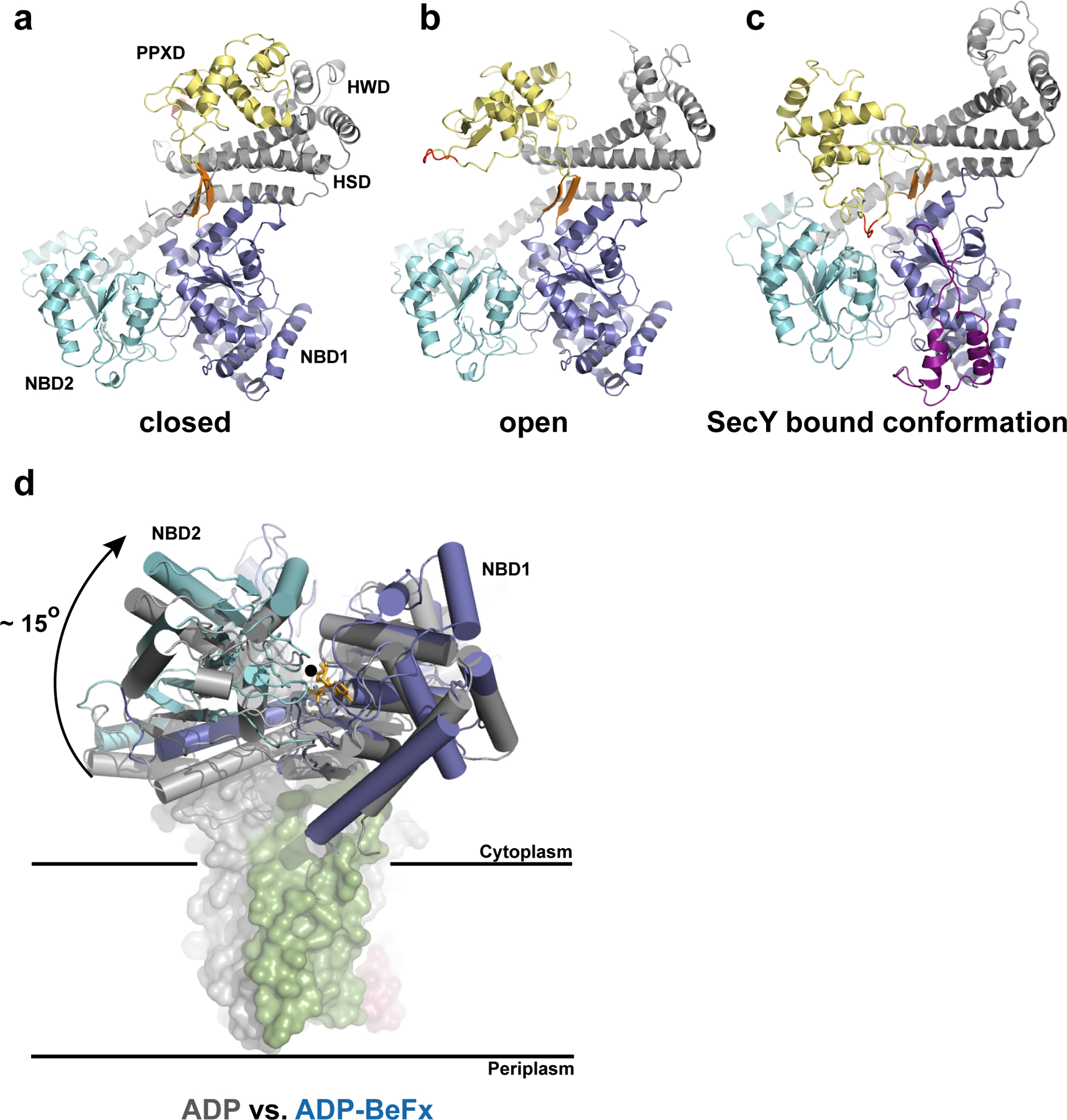
Figure 2.

Conformational changes of SecA. a–c, Different orientations of the PPXD relative to the NBDs. The view is from the cytosol, as in Fig. 1c. The closed (a) and open (b) conformations refer to the B. subtilis SecA structures (PDB accessions 1M6N and 1TF2, respectively). The loop of the PPXD contacting NBD1 and NBD2 in the SecA–SecY complex is highlighted in red. The insertion in the T. maritima NBD1 is shown in purple. The conserved β-strands connecting NBD1 with the PPXD are shown in orange. d, Position of the NBDs in the ADP-bound state of B. subtilis SecA (grey) versus the ADP–BeF x bound state of T. maritima SecA (blue) when associated with SecY. The structures were aligned with respect to NBD1. The rotation axis indicated by a black circle is parallel to the plane of the membrane. The modelled ADP–BeF3-is shown as orange sticks.
