
Fig. 6. A. muciniphila–induced pTregs prevent colitis in the adoptive transfer model.
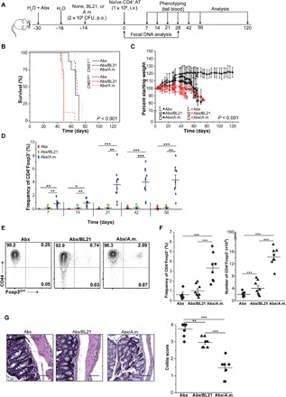
(A) Outline of the experiment. Mice were treated with a cocktail of antibiotics (Abx) and inoculated with indicated bacteria (n = 8 for each condition). At indicated time points, tail blood and feces samples were collected. i.v., intravenously. (B) Survival curves of adoptive transfer recipients precolonized with indicated bacteria (Kaplan-Meier curves and log-rank test). (C) Loss of initial body weight following adoptive transfer. Each point represents the mean percentage of initial body weight for the cohort ± SEM. (D) Frequency of pTregs in the blood at indicated time points for CNS1+/+ cell recipients. (E) Representative staining of analysis of colonic CD4+CNS1+/+ cells in recipient mice. (F) Proportions and total number of colonic CD4+ cells in individual mice after colonization and adoptive transfer. (G) Histological analysis of colonic sections. Representative H&E staining (×40 magnification) and colitis scores (scores are presented as an average of three fragments of each tissue) are shown. Scale bars, 100 μm. The experiment was repeated twice with three to five mice per group. Each symbol depicts individual animal. For survival analysis, the log rank test was performed; otherwise, paired t tests were applied; *P < 0.05, **P < 0.01, ***P < 0.001. BL21-E. coli BL21DE3.