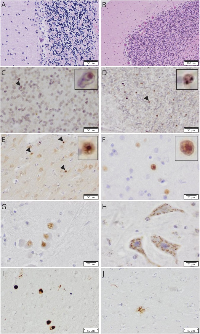
Figure 2. Histopathologic features of affected cases III-13 (A–H) and III-10 (I and J).
(A and B) Nearly complete loss of Purkinje cells, loss of neurons, and spongiosis in the cerebellar granular layer of the patient (A) compared with the control brain (B) in H/E staining; (C and D) NII in the cerebellar granular layer with ubiquitin (C) and p62 staining (D); (E) NII in the occipital cortex with p62 staining; (F) DNS in the occipital cortex with 1C2 staining; (G) DNS in the inferior olive with 1C2 staining; (H) granular CI in the hypoglossal nucleus with 1C2 staining; (I) AT8-positive neurons, neurites, and glial staining in the thalamic nuclei; (J) AT8-positive tufted astrocyte in the putamen. The arrowheads indicate NII, which are magnified in figure 2C-F. CI = cytoplasmic inclusion; DNS = diffuse nuclear staining; H/E = hematoxylin and eosin staining; NII = neuronal intranuclear inclusion.

