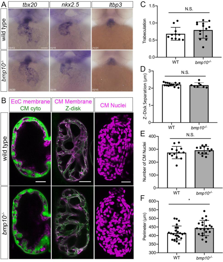
Fig. 3. Embryonic ventricular myocardium development is unaffected bmp10 mutants.
a Whole-mount in situ hybridization for tbx20, nkx2.5, and ltbp3 at 2 dpf, wild type and bmp10pt527 mutant siblings. Ventral views, anterior top. Numbers in left-hand corner show number of embryos with shown phenotype over total number of embryos imaged over 3 experiments. Scale bar: 100 μm. b Ventricle morphology at 5 dpf in wild type and bmp10pt527 mutant siblings. Left panels, trabeculation: Tg(fli1a.ep:mRFP-CAAX)pt505;Tg(myl7:EGFP)twu34, endocardial cell (EcC) membranes magenta, cardiomyocyte (CM) cytoplasm green. Middle panels, sarcomeric architecture: Tg(myl7:mKATE-CAAX)sd11;Tg(myl7:actn3b-EGFP)sd10; CM membranes magenta, Actn3b (Z-disks) green. Right panels, CM number: Tg(myl7:dsRed2-NLS)f2; CM nuclei magenta. Confocal images, single planes (left, middle) or 2D projections (right). Scale bars: 25 μm. c Trabeculation [(trabecular trace-central chamber trace)/central chamber trace)] at 5 dpf, N = 11 wild types, 14 mutants over 4 experiments. d Z-disk separation, N = 17 wild types, 8 mutants over 5 experiments. e Number of CM nuclei, N = 13 wild types, 13 mutants over 4 experiments. f Ventricle outer perimeter, N = 24 wild type, 22 mutants over 8 experiments. Bars represent mean ± SD. Unpaired t-test, *P < 0.05, NS = not significant.

