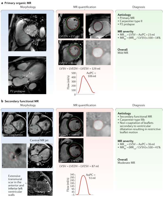
Fig. 3. Case studies of primary and secondary MR.
a | Mitral regurgitation (MR) assessment with cardiovascular magnetic resonance imaging in a patient with organic MR. Prolapse of the P2 can be seen on the three-chamber view during mid-systole (morphology panel, bottom image), and the resulting MR jet is visualized on the short-axis view (morphology panel, top image). The MR volume (MRvol) is quantified using the standard method: left ventricular stroke volume (LVSV) minus aortic phase-contrast forward volume (AoPC). b | MR assessment in a patient with ischaemic cardiomyopathy. Non-coaptation owing to ventricular dilatation is seen on the short-axis cines (morphology panel, top images). A through-plane phase-contrast acquisition shows the central MR jet (morphology panel, right-hand middle image). Late gadolinium enhancement imaging reveals extensive ischaemic myocardial scaring (Morphology panel, right-hand bottom image). LVEDV, left ventricular end-diastolic volume; LVESV, left ventricular end-systolic volume; MRRF, mitral regurgitation fraction.

