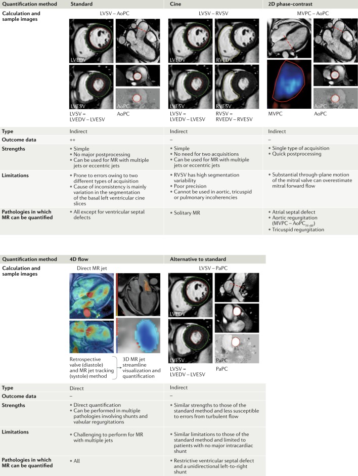
Fig. 5. Main methods of MR quantification by cardiovascular magnetic resonance imaging.
Prognostic and diagnostic outcome data are most available for the standard method of quantifying mitral regurgitation (MR) volume (MRvol), which is left ventricular stroke volume (LVSV) minus aortic phase-contrast forward volume (AoPC). Other methods have particular advantages or disadvantages. In routine clinical practice, cross-checking between methods is recommended. FF+BF, forward flow plus backward flow; LVEDV, left ventricular end-diastolic volume; LVESV, left ventricular end-systolic volume; MVPC, mitral valve phase-contrast stroke volume; PaPC, pulmonary artery phase-contrast stroke volume; RVEDV, right ventricular end-diastolic volume; RVESV, right ventricular end-systolic volume; RVSV, right ventricular stroke volume.

