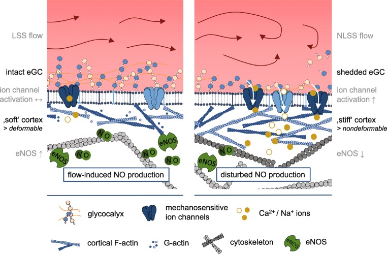
Fig. 2.
Model of eGC- and ion channel-mediated mechanosignaling. Physiological LSS is accompanied by an intact eGC structure and a “soft” and deformable actin cortex. EC can react to changes in blood flow with increased eNOS activity and NO-mediated vasodilation (left figure). Pathophysiological increase of shear stress (e.g., by NLSS) leads to a disturbed eGC structure, increased Ca2+, and Na+ influx and stiffening of the cell cortex. This is accompanied by reduced eNOS activity and impaired flow-mediated vasodilation (right figure). The ability of the EC to change their mechanical properties, i.e., to alternate between “stiff” and “soft” conditions, is an important physiological feature. Loss of this plasticity leads to a dysfunctional endothelium

