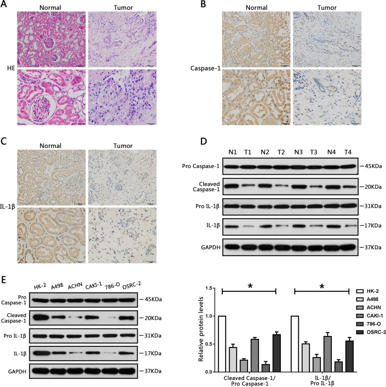
Fig. 1. Downregulation of Caspase-1 and IL-1β in RCC tissue specimens and cell lines.
a Hematoxylin and eosin staining of renal cell carcinoma (RCC) specimens and adjacent normal renal tissues. b Immunohistochemical staining of caspase-1 in RCC specimens and adjacent normal renal tissues. c Immunohistochemical analysis of IL-1β in RCC tissues and adjacent normal renal tissues. d Western blot analysis of expression levels of caspase-1 and IL-1β in RCC tissues and surrounding normal regions. e Relative caspase-1 and IL-1β protein levels in HK-2 cells (human renal tubule epithelial cell line), and five RCC cell lines (A498, ACHN, CAKI-1, 786-O, and OSRC-2) were analyzed using western blot analysis. Results are shown as means ± SD. *P < 0.05, relative to the HK-2 cell line.

