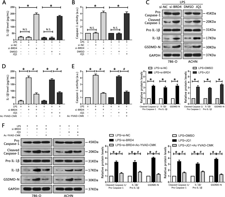
Fig. 3. BRD4 inhibition activated caspase-1-dependent pyroptosis in RCC cell lines.
a 786-O and ACHN cells were pretreated with LPS though the methods mention above, then transfected with siBRD4 or treated with JQ1, respectively, and the levels of IL-1β secreted into the culture medium were assessed using ELISA. b Activity of caspase-1 following siBRD4 transfection and JQ1 treatment was measured. c Western blots of caspase-1, IL-1β and GSDMD levels in 786-O and ACHN cells following transfection with siBRD4 or treatment with JQ1, respectively. The bar graph shows the relative levels of caspase-1, IL-1β and GSDMD from three independent experiments. d–f 786-O and ACHN cells were treated with 50 μM Ac-YVAD-CMK for 24 h, then transfected with siBRD4 or treated with JQ1. d Production of IL-1β secreted into the culture medium was measured using ELISA. e Caspase-1 activity was evaluated in 786-O and ACHN cells. f Western blot analysis of caspase-1, IL-1β and GSDMD levels. The bar graph shows the relative levels of caspase-1 and IL-1β from three independent experiments. *P < 0.05 vs. control; #P < 0.05 vs. si-BRD4 or JQ1; N.S. P > 0.05 vs. control.

