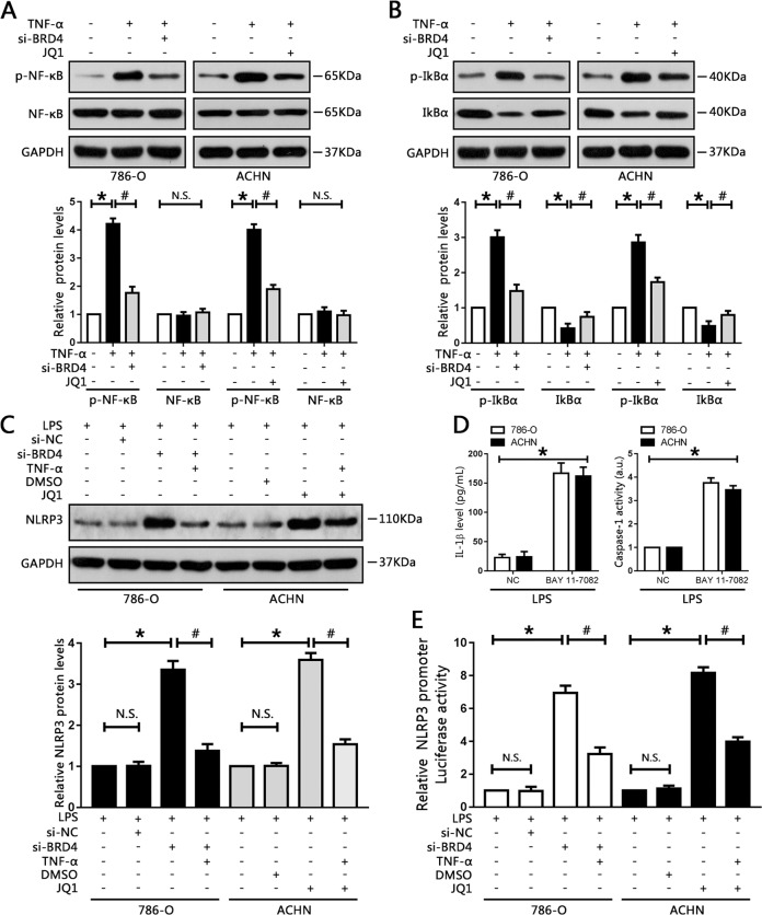
Fig. 7. BRD4 regulated NLRP3 expression via the NF-κB pathway.
786-O and ACHN cells were pre-treated with 10 ng/ml TNF-α for 1 h before administration of si-BDR4 or JQ1, respectively. a, b Nuclear factor κB, phosphorylated NF-κB, IκBα, and phosphorylated IκBα protein levels were measured by western blotting, and quantified. *P < 0.05 vs. control. #P < 0.05 vs. TNF-α. c Relative NLRP3 protein levels were measured by western blot analysis, and quantified. *P < 0.05 vs. control; #P < 0.05 vs. si-BRD4 or JQ1; N.S. P > 0.05 vs. control. d Following treatment with 10 μM BAY 11-7082 for 1 h, levels of IL-1β secreted into the cell culture supernatants, and caspase-1 activity, were measured. *P < 0.05 vs. NC. e NLRP3 promoter activity in the indicated groups was measured using luciferase assay. Experiments were performed in triplicate. *P < 0.05 vs. control; #P < 0.05 vs. si-BRD4 or JQ1; N.S. P > 0.05 vs. control.

