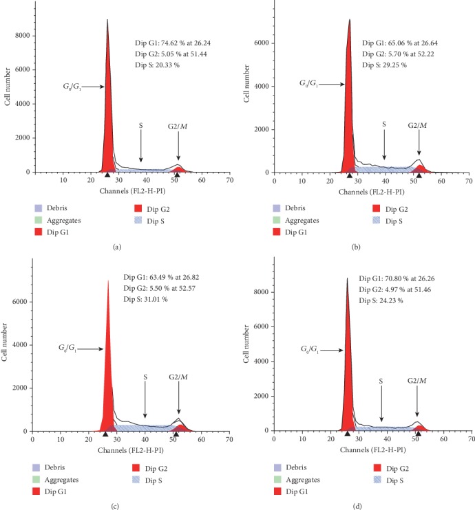
Figure 3.

Representative images of PC3 cell cycle analysis. (a) Control group (PC3 cells), (b) PC3 cells cocultured with hBMSC cells, (c) PC3 cells cocultured with hFOB1.19 cells, and (d) PC3 cells cocultured with hFOB1.19 cells+scutellarin. The ordinate shows the number of cells counted, and the abscissa shows DNA content. G2/G1 was 2.0 (i.e., the cells were tetraploid in the G2 phase and diploid in the G1 phase, with a ratio of 2).
