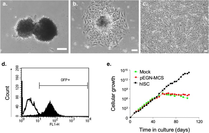
Fig. 1.
Isolation and selection of immortalized islet-derived cells. Phase-contrast microscopic view of pancreatic islets handpicked after dithizone staining (a, day 1). Islets adhered and cells spread-out (b, day 3) until becoming a monocellular layer of fibroblast-like cells (c, day 40). Scale bar = 100 μm. Efficiency of hTERT retroviral infection was assessed by neomycin resistance and GFP expression. GFP-positive cells were individualized by flow cytometry (d). GFP-sorted cells, defined as hISCs, did not shown senescence (black line) while non-infected cells (Mock, green line) or empty vector-transduced cells (pEGN-MCS, red line) stopped proliferating after 50 days (e)

