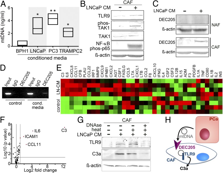
Fig. 1.
Activation of TLR9 and C3a by mtDNA. (A) mtDNA was measured from conditioned medium (CM) of prostatic epithelia after 48 h of incubation (n = 3). (B) Protein expression in CAF treated with LNCaP-CM was visualized by Western blot. (C) DEC205 expression was measured in NAF and CAF treated with LNCaP-CM by Western blot. (D) DEC205 was immunoprecipitated, cross-linked, and subjected to mtDNA PCR amplification for MT-CO2 following CAF incubation with fresh RPMI or LNCaP-CM. CAF cell lysate prior to immunoprecipitation or IgG immunoprecipitant was used as total input and negative controls, respectively. (E) mRNA expression profiles of the NF-κB–signaling targets in CAF incubated with LNCaP-CM were compared to control-CM in the heat map (n = 4). (F) Volcano plot showing distribution of differential mRNA expression levels in CAF and CAF incubated with LNCaP-CM. While only secreted proteins were illustrated in the heat map, all 84 NF-κB target genes were represented in the volcano plot. (G) TLR9 and anaphylatoxin C3a protein expression was visualized in CAF incubated with LNCaP-CM incubated with or without DNase1 treatment. DNase activity was heat inactivated after 10 min. (H) MtDNA secreted by PCa epithelia signal CAF by binding DEC205 and subsequent TLR9-mediated anaphylatoxin C3a expression. *P < 0.05, **P < 0.01.

