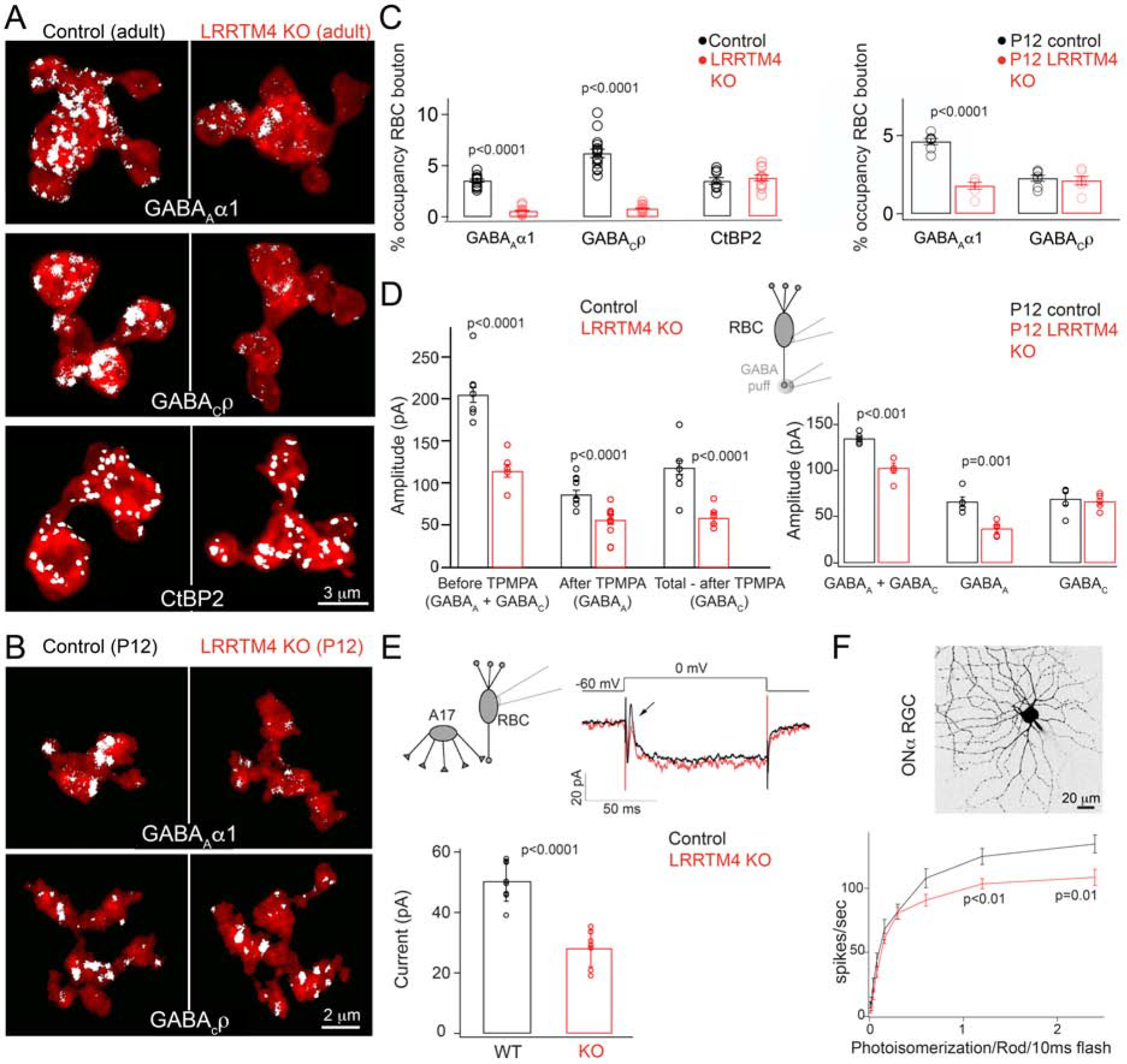
Figure 2: Absence of LRRTM4 perturbs GABAergic synapses on retinal rod bipolar axon terminals.

(A) White pixels represent signal above background of immunolabeled GABAAα1 and GABACρ receptors and the ribbon marker CtBP2 within RBC terminals. Shown are examples from adult littermate control (Control) and LRRTM4 KO retina. (B) GABAAα1 and GABACρ receptor expression on P12 RBC axons of control and LRRTM4 KO. (C) Left: Quantification of percent (%) occupancy of GABAAα1, GABACρ and CtBP2 within adult RBC. Right: % occupancy of GABAAα1 and GABACρ within P12 RBC boutons. (D) Peak response amplitudes evoked by a puff of GABA at the RBC terminal during whole cell voltage-clamp recording of RBCs. The GABAC receptor antagonist TPMPA was used to separate the GABAA and GABAC mediated response components. Left: Adult RBCs, number of cells: 12 for control and 11 for KO. Right: P12 RBC responses, number of cells: 5 for control and KO. (E) Exemplar trace of the step evoked inhibitory A17 feedback current measured from adult RBCs in control and LRRTM4 KO retinas. Arrow points to the A17 mediated feedback current. Bar plot of the feedback current amplitude across genotypes. Number of cells: 8 control and 7 KO. (F) Cell-attached recordings of adult ON alpha GCs to measure spike responses to increasing flash strengths at dim-light levels. Top: Image of an ON alpha GC. Bottom: Spike rates of ON alpha GC in response to increasing flash strengths in control and LRRTM4 KO retinas. Number of recorded cells: 7 for control and 9 for KO. Significant differences are indicated by p-values; two-tailed unpaired T-test between genotypes. For each of panels C-F, N≥3 pairs of littermate control-KO animals were used.
