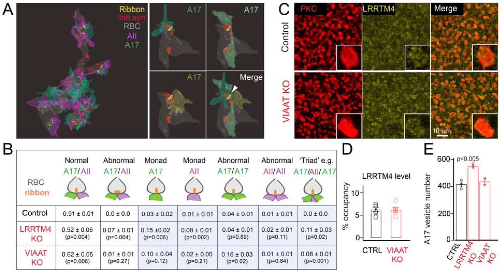
Figure 4: Impaired inhibitory input onto rod bipolar cell axons results in structural mis-arrangements of ribbon synapses.

(A) Left: 3D view of the RBC terminal in the VIAAT KO retina. Ribbons, inhibitory synapses and A17 and AII partner profiles are shown. Right: Example of a RBC ‘triad’ ribbon synapse with three A17 partners (white arrowhead) in the VIAAT KO retina. (B) Table summarizing adult RBC dyad arrangements from reconstructions across genotypes. N=3 Control, 3 LRRTM4 KO and 2 VIAAT KO RBCs from 2 pairs of littermate control and KO animals. The numbers depict the average fraction (mean ± S.E.M) of ribbon synapses with each type of illustrated structural arrangement. In controls, 0.01 ± 0.01 ribbons were also found juxtaposed to a single AII or an A17 partner, and an unidentified, presumably ganglion cell partner (not included in summary table). Triad configurations include an AII and two A17 partners (illustrated here), three A17 partners or two AII with one A17 partner. P values listed for two-tailed unpaired T-test between control and LRRTM4KO, and between control and VIAAT KO. Abnormal monad, dyad and triad arrangements are enclosed within blue boxes and include both vertical and horizontal ribbon arrangements. (C) LRRTM4 expression at PKC-immunopositive RBC terminals appears normal in the VIAAT KO retina. (D) Quantification of the percent (%) occupancy of LRRTM4 within adult RBC boutons of control (CTRL) and VIAAT KO retinas is comparable. (N>3 animal pairs). (E) Quantification of synaptic vesicle number within A17 boutons in CTRL, LRRTM4 KO and VIAAT KO (N=3 Control, 3 LRRTM4 KO and 2 VIAAT KO from 2 pairs of littermate control and KO animals; for each RBC 10 A17 boutons were averaged). P value listed for two-tailed unpaired T-test between control and LRRTM4 KO.
