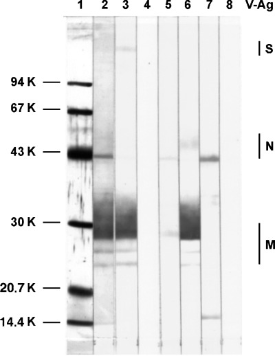
Figure 1.

Western blot analysis of TGEV antibodies. After separation in 12% polyacrylamide gel, purified TGEV and a low molecular weight standard (LMW) were transferred to a nitrocellulose membrane. A part of the membrane with the LMW standard was stained with colloidal gold (lane 1). The other lanes with TGEV were incubated with swine antisera to TGEV (lanes 2 and 3), mAbTGEV D3/G6 (lane 5), D7/G7 (lane 6), B7/F7 (lane 7) or with diluting solution alone (lanes 4 and 8). After incubation with peroxidase conjugates to swine (lanes 2–4) and mouse (lanes 5–8) immunoglobulins, the reaction was visualised by incubation in a substrate solution containing chromogen DAB. The localization of TGEV antigens S, N and M is indicated.
