
Figure 2.
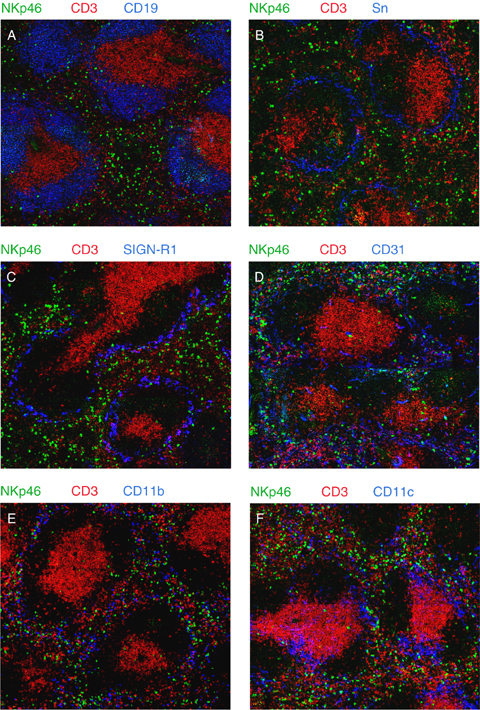
Localization of natural killer (NK) cells in the spleen. Frozen sections of spleen were fixed with acetone and stained with fluorescently coupled antibodies or biotinylated antibodies revealed with fluorochrome‐coupled streptavidin. Anti‐CD3 (145‐2C11), anti‐CD19 (1D3), anti‐CD31 (MEC13‐3), anti‐CD11b (M1/70), and anti‐CD11c (HL3) monoclonal antibodies were from BD Pharmingen (San Diego, CA, USA). Anti‐sialoadhesin (MOMA‐1 for metallophilic macrophages) and anti‐Sign‐R1 (ER‐TR9 for marginal zone macrophages) monoclonal antibodies were obtained from AbD Serotec (Raleigh, NC, USA) and BMA Biomedicals (Augst, Switzerland), respectively. Polyclonal goat anti‐NKp46 (R&D Systems, Minneapolis, MN, USA) was revealed with donkey‐anti‐goat antibody (Invitrogen, Carlsbad, CA, USA), Sections were visualized by confocal microscopy (Zeiss LSM 510 META, Iena, Germany). Panels A–F show representative images for the indicated staining.