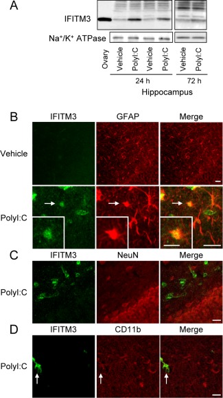
Figure 1.

IFITM3 proteins are expressed predominantly in astrocytes, but not neurons and microglia, in vivo. (A) Hippocampal IFITM3 protein levels at 24 h and 72 h after the final polyI:C treatment in neonatal mice. An ovary sample was loaded as a positive control. (B–D) Neonatal polyI:C treatment increases IFITM3‐like immunoreactivity in astrocytes. Double immunostaining for IFITM3 (green in left panels: B–D), GFAP (red in middle panels: B), NeuN (C), and CD11b (D), in the hippocampus at 24 h after the final polyI:C treatment of neonatal mice. Scale bar: 20 µm (inset, 10 μm).
