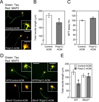
Figure 3.

Effect of polyI:C‐ACM on cell viability or neurite elongation of cultured neurons. (A) Immunocytochemical images of primary neurons cultured for 24 h from DIV2 with control‐ or polyI:C‐ACM derived from ICR mice. Neurons were fixed and stained with tau (green: axonal marker) and MAP2 (red: dendritic marker) antibodies at 24 h after treatment with control‐ or polyI:C‐ACM. Scale bar: 200 µm (inset, 75 μm). (B) Total neurite length of each neuron. Values are the means ± SEM of three independent experiments (n = 72–75). *P < 0.05 versus control‐ACM. (C) Cell viability of primary cultured neurons was assayed at 24 h after polyI:C‐ACM treatment by MTT assays. Values are the means ± SEM (n = 5). (D) Immunocytochemical images of neurons cultured with control‐ or polyI:C‐ACM derived from wt (C57BL/6J) or ifitm3 −/− astrocytes. Scale bar: 200 µm (inset, 100 μm). (E) Total neurite length of each neuron. Values are the means ± SEM of three independent experiments (n = 25) [polyI:C‐ACM (F (1,90) = 9.86, P < 0.001), genotype (F (1,90) = 0.05, P = 0.82), interaction (F (1,90) = 0.075, P = 0.78)]. *P < 0.05 versus wt/control‐ACM, # P < 0.05 versus wt/polyI:C‐ACM.
