
Figure 7.
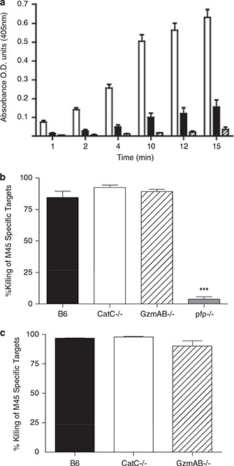
Antiviral CTL function is normal in CatC mice. (a) B6 mice (open bar), B6.CatC−/− (solid bar) and GzmAB−/− (hatched bar) mice were infected with MCMV and CD8+ T cells purified at day 6 p.i. Lysates prepared from these cells were assayed for GzmB activity by measuring the hydrolysis of peptide substrate. Data are pooled from two independent experiments (n=4). (b) The indicated mouse strains were infected with 5 × 10³ PFU of MCMV‐K181‐Perth. At day 6 p.i., mice were injected intravenously with a 1:1 mixture of CFSE‐labeled M45‐pulsed targets cells and nonpulsed targets. A single‐cell suspension from the spleens of the infected mice was prepared 4 h after target cell transfer and loss of peptide‐pulsed targets measured by FACS analysis. B6 mice (solid bar), B6.CatC−/− (open bar), B6.GzmAB−/− (hatched bar) and B6.Pfp−/− (grey bar) mice. Data from two independent experiments have been pooled, mean±s.e.m. are plotted (n⩾8). ∗∗∗ P=0.0004. (c) Mice were infected with MCMV and peptide‐labeled target cells injected as described. A single‐cell suspension was prepared from livers of infected mice 16 h after target cell transfer and loss of peptide pulsed targets measured by FACS analysis. B6 mice (solid bar), B6.CatC−/− (open bar), B6.GzmAB−/− (hatched bar).