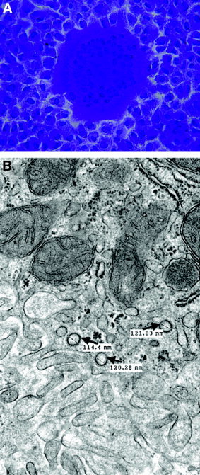
Figure 3.

Assays for detection of coronavirus infection (MHV). (A) Plaque assay with fusion of cells (syncytia) on a monolayer of L2 cells stained with crystal violet. (B) Electron micrograph of liver at 3 days after infection showing typical MHV virions in a hepatocyte (arrow). The virions are round, mildly pleomorphic (100–120 nm), and show surface membrane thickening indicative of the corona peplomer structure.
