Fig. 3.
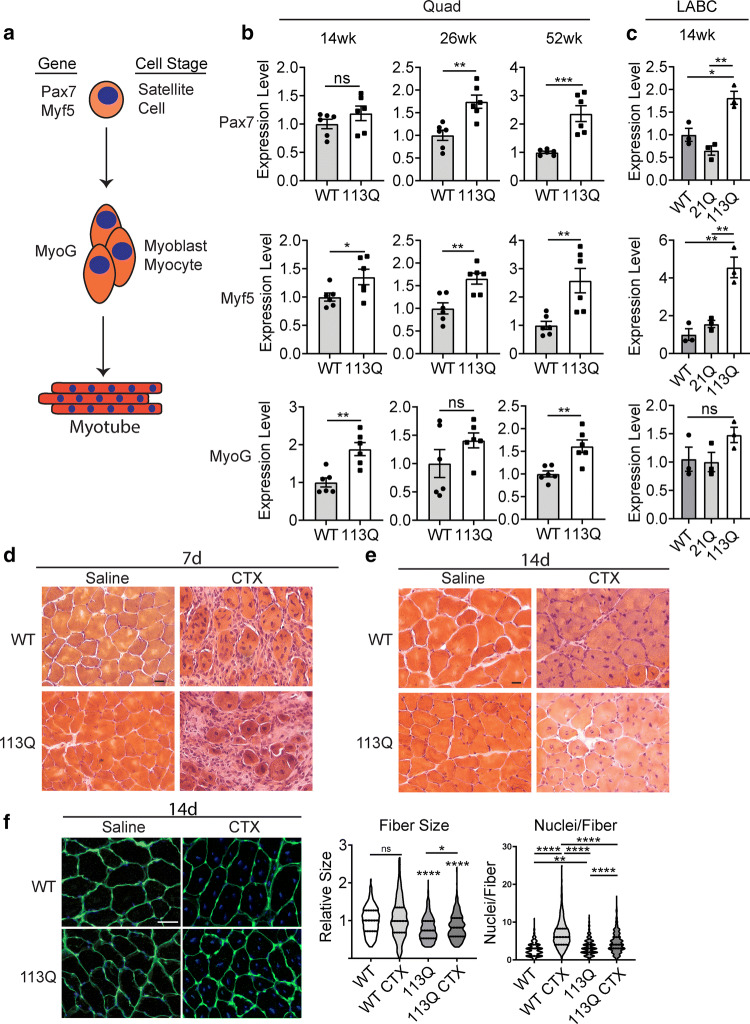
Myoregeneration is intact in AR113Q muscle. a Schematic of examined markers of muscle satellite cells (PAX7, Myf5) and myoblasts/myocytes (MyoG). b, c Relative mRNA expression of PAX7 (top), Myf5 (middle) and MyoG (bottom) in WT and AR113Q quadriceps at 14, 26, and 52 weeks (b, n = 6/group) and in WT, AR21Q, and AR113Q LABC at 14 weeks (c, n = 3/group). d–f Tibialis anterior of WT or AR113Q mice injected with cardiotoxin (CTX) or saline at 19 weeks and examined by H&E stain at 7 days (d) or 14 days (e) post-injection. Scale bar = 20 µm. (f, Left) Muscle fibers were stained with Wheat Germ Agglutinin (WGA) to delineate muscle fiber membranes and DAPI at 14 days post-injection. Fiber size and nuclei/fiber were quantified (f, right) at 14 days post-injection and are displayed as a violin plot (n = 3 mice/group, 3 images/mouse, > 100 fibers/mouse). Scale bar = 50 µm. ns not significant (p > 0.05), *p < 0.05, **p < 0.01, ***p < 0.001, ****p < 0.0001 by unpaired t test (b), one-way ANOVA with Tukey’s post hoc test (c), and two-way ANOVA with Tukey’s post hoc test (f, left and right panel)

