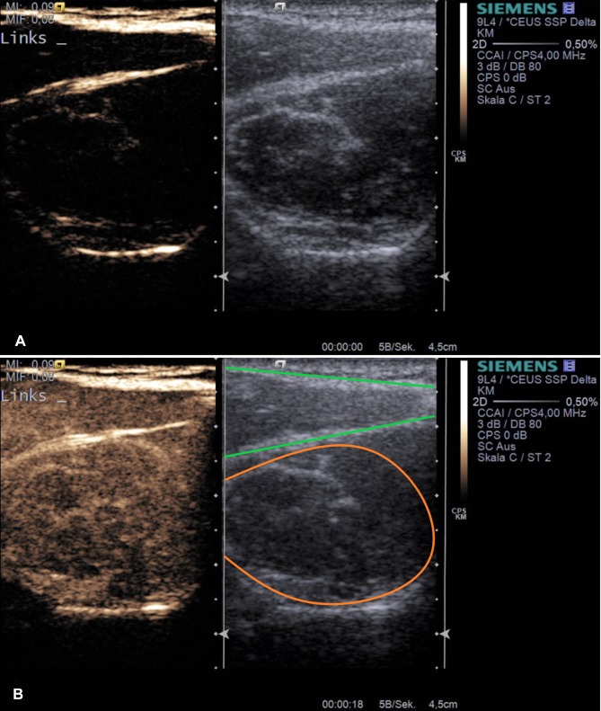
Figure 1.
Example application of musculoskeletal CEUS. Dual modes with combined Cadence™ contrast mode and conventional B-mode ((A) before contrast agent application) are displayed after supraspinatus tendon tear (muscular area) inside the supraspinatus fossa (a+b). The lower image (B) represents the corresponding image after sulfur hexafluoride contrast agent application with maximum enhancement. Orange – supraspinatus muscle, green – trapezius muscle.

