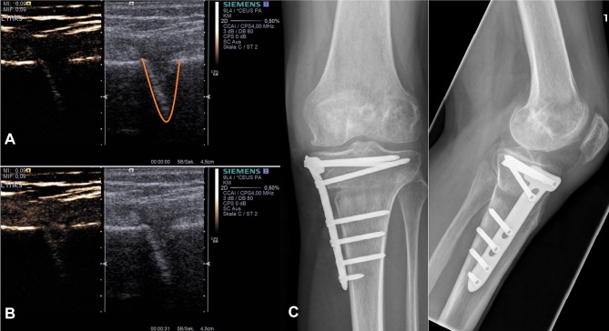
Figure 3.
Example application of skeletal osteotomy CEUS. Dual modes with combined Cadence™ contrast mode ((A) before contrast agent application) and conventional B-mode (a+b) are displayed after HTOWO (C). The lower image (B) represents the corresponding image after sulfur hexafluoride contrast agent application with maximum enhancement. Right images show the corresponding X-ray of the patient (C). Orange – osteotomy gap.

